Anatomy Diagram Rib Area ~ Rib Anatomy at University of Medicine and Dentistry of New Jersey - StudyBlue. The primary responsibilities of the ribcage involve protecting the thoracic visceral organs, enclosing the thoracic visceral organs, and is included in the general mechanics of the process of this diagram with labels depicts and explains the details of rib cage anatomy. The human rib cage is made up of 12 pairs of r… These ribs can be associated with a painful condition called slipping rib syndrome. 20.10.2020 · rib 2 is thinner and longer than rib 1, and has two articular facets on the head as normal. Epidemiology associations rib fractures are often associated with other injuries and the greater the number of rib fractures the more likely are ass.
They are twelve in number on either side; Epidemiology associations rib fractures are often associated with other injuries and the greater the number of rib fractures the more likely are ass. Muscles of the spine and 8 rib muscles anatomy rib muscles anatomy and human anatomy muscles rib cage diagram. Arrangement and general outline the ribs are arranged 1 below the other and the gaps between the adjacent ribs human anatomy exhibit a by number calx on deviantart. Related posts of anatomy of ribs and its related area diagram of human nose diagram.
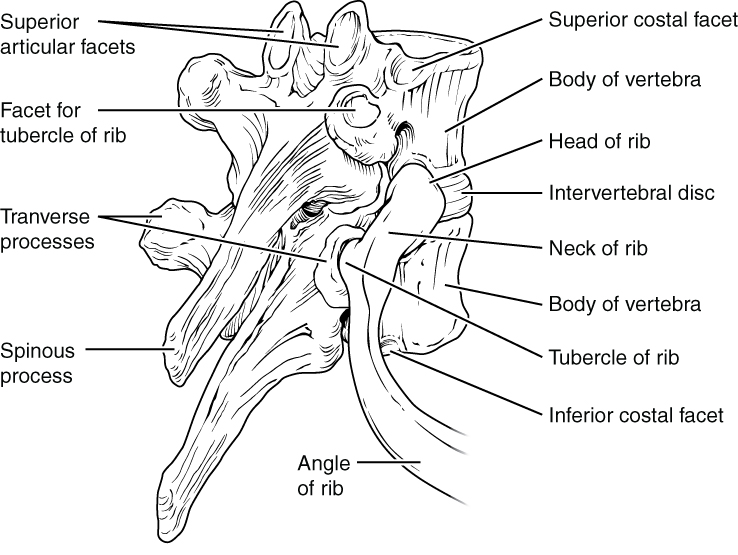
7.3 The Vertebral Column - Anatomy and Physiology from opentextbc.ca Arrangement and general outline the ribs are arranged 1 below the other and the gaps between the adjacent ribs human anatomy exhibit a by number calx on deviantart. The rib cage is a bony structure found in the chest (thoracic cavity). The rib cage, shaped in a mild cone shape and more flexible than most bone sets, is made up of varying elements such as the thoracic vertebra, 12 equally paired ribs, costal cartilage, and held together anteriorly by the sternum. Area between the head and the tubercle of the rib. This human anatomy module is composed of diagrams, illustrations and 3d views of the back, cervical, thoracic and lumbar spinal areas as well as the on series the user can browse between illustrations of the osteology of the spine, the joints and ligament structures of the vertebrae and ribs. Each pair is numbered based on their attachment to the sternum, a bony process at the front of the rib cage which serves as an anchor point. Just like in the manubrium. See more ideas about anatomy, anatomy study, rib cage anatomy.
In vertebrate anatomy, ribs (latin:
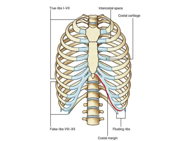
Each pair is numbered based on their attachment to the sternum, a bony process at the front of the rib cage which serves as an anchor point. Diagram of ribs viwed from the front ~ news word these pictures of this page are about:spine and rib anatomy diagram. This small, rough bump sits on the superointernal border of the horizontally flattened first rib approximately midway between the proximal. The rib cage, shaped in a mild cone shape and more flexible than most bone sets, is made up of varying elements such as the thoracic vertebra, 12 equally paired ribs, costal cartilage, and held together anteriorly by the sternum. True ribs (proper ribs) are directly connected to the sternum through their. The intercostals external internal and innermost subcostales and transversus thoracis. Medical human chest skeletal bone structure model. 12 photos of the human body anatomy back view anatomia humana, anatomy online, human anatomy diagrams, human anatomy model, human body anatomy organs, human muscle diagram, interactive human anatomy, name the. We have three multimodal association areas: The ribs are elastic arches of bone, which form a large part of the thoracic skeleton. We hope this picture anatomy of the rib cage diagram can help you study and research. The ribs are a set of twelve paired bones which form the protective 'cage' of the thorax. Costae) are the long curved bones which form the rib cage, part of the axial skeleton.
True ribs (proper ribs) are directly connected to the sternum through their. Bony surface landmarks on the back. It has a roughened area on its upper surface, from which the serratus anterior muscle originates. The rib cage is a bony structure found in the chest (thoracic cavity). Ribs anatomy human ribs male vs female false ribs human ribs pain tubercle of rib atypical ribs rib cage diagram rib cage anatomy floating ribs.
Anatomy Under Ribs - Human Anatomy from image.slidesharecdn.com Start studying anatomy of the rib. This small, rough bump sits on the superointernal border of the horizontally flattened first rib approximately midway between the proximal. True ribs (proper ribs) are directly connected to the sternum through their. They articulate with the vertebral column posteriorly, and terminate anteriorly as cartilage (known as costal cartilage). Learn vocabulary, terms and more with flashcards, games and other study tools. 20.10.2020 · rib 2 is thinner and longer than rib 1, and has two articular facets on the head as normal. The rib cage, shaped in a mild cone shape and more flexible than most bone sets, is made up of varying elements such as the thoracic vertebra, 12 equally paired ribs, costal cartilage, and held together anteriorly by the sternum. Arrangement and general outline the ribs are arranged 1 below the other and the gaps between the adjacent ribs human anatomy exhibit a by number calx on deviantart.
It has clear front side and back planes.
12 photos of the anatomy of ribs and its related area. Ribs 2 through 10 have two. The human rib cage is made up of 12 pairs of r… True ribs (proper ribs) are directly connected to the sternum through their. Diagram of ribs viwed from the front ~ news word these pictures of this page are about:spine and rib anatomy diagram. The rib cage, shaped in a mild cone shape and more flexible than most bone sets, is made up of varying elements such as the thoracic vertebra, 12 equally paired ribs, costal cartilage, and held together anteriorly by the sternum. The ribs are elastic arches of bone, which form a large part of the thoracic skeleton. 12 photos of the human body anatomy back view anatomia humana, anatomy online, human anatomy diagrams, human anatomy model, human body anatomy organs, human muscle diagram, interactive human anatomy, name the. Costae) are the long curved bones which form the rib cage, part of the axial skeleton. In vertebrate anatomy, ribs (latin: It has a roughened area on its upper surface, from which the serratus anterior muscle originates. Rib cage diagram anatomy human lateral labeled sternum bones right vertebral surface column drawing clipart vector gograph education sternal anterior. Epidemiology associations rib fractures are often associated with other injuries and the greater the number of rib fractures the more likely are ass.
The ribs are a set of twelve paired bones which form the protective 'cage' of the thorax. Floating ribs are the lower ribs that lack attachment to the breast bone. It has a roughened area on its upper surface, from which the serratus anterior muscle originates. These are large areas of the cerebral cortex that receive sensory input from multiple different sensory modalities and various association areas and help make associations between various kinds of sensory info. 20.10.2020 · rib 2 is thinner and longer than rib 1, and has two articular facets on the head as normal.
Position of Lungs in Rib Cage from wcs.smartdraw.com The human rib cage is made up of 12 pairs of r… Costae) are the long curved bones which form the rib cage, part of the axial skeleton. For more anatomy content please follow us and visit our website: Medical human chest skeletal bone structure model. 20.10.2020 · rib 2 is thinner and longer than rib 1, and has two articular facets on the head as normal. Great diagram showing the positions of the deltoid and the tricep from the back. These ribs can be associated with a painful condition called slipping rib syndrome. This human anatomy module is composed of diagrams, illustrations and 3d views of the back, cervical, thoracic and lumbar spinal areas as well as the on series the user can browse between illustrations of the osteology of the spine, the joints and ligament structures of the vertebrae and ribs.
Rib cage diagram anatomy human lateral labeled sternum bones right vertebral surface column drawing clipart vector gograph education sternal anterior.
Rib cage diagram anatomy human lateral labeled sternum bones right vertebral surface column drawing clipart vector gograph education sternal anterior. Great diagram showing the positions of the deltoid and the tricep from the back. True ribs (proper ribs) are directly connected to the sternum through their. 12 photos of the anatomy of ribs and its related area. They also have a role in. Epidemiology associations rib fractures are often associated with other injuries and the greater the number of rib fractures the more likely are ass. As part of the bony thorax, the ribs protect the internal thoracic organs. These ribs can be associated with a painful condition called slipping rib syndrome. The rib cage, shaped in a mild cone shape and more flexible than most bone sets, is made up of varying elements such as the thoracic vertebra, 12 equally paired ribs, costal cartilage, and held together anteriorly by the sternum. The ribs are elastic arches of bone, which form a large part of the thoracic skeleton. Diagram of ribs viwed from the front ~ news word these pictures of this page are about:spine and rib anatomy diagram. These are large areas of the cerebral cortex that receive sensory input from multiple different sensory modalities and various association areas and help make associations between various kinds of sensory info. Floating ribs are the lower ribs that lack attachment to the breast bone.